
Unmasking Skin Lesions
Skin lesions can be a cause for concern, but some can be benign. To navigate through the complexities of skin health, medical professionals often rely on specific criteria and rules. In this article, we'll dive into two widely-used methodologies: the 7 Points Checklist (7PCL) and the ABCDE rule, both designed to help identify potential risks in skin lesions.
Weighted scoring with 7 Points Checklist (7PCL)
The 7PCL, a method developed in the 1980s to detect cancerous lesions (mainly melanoma), has now evolved as a structured approach for evaluating skin lesions, assigning weighted scores based on various signs. Here's a breakdown of the scoring system:
Major Signs (2 points each):
- Change in size of lesion
- Irregular pigmentation: Variation in pigmentation, especially irregular patterns or networks
- Irregular border: Borders that are not well-defined
Minor Signs (1 point each):
- Inflammation: Inflammatory reactions may hint at underlying problems within the lesion.
- Crusting/Bleeding: Lesions that exhibit crusting or bleeding could be undergoing abnormal processes.
- Any alterations in sensory perception
- Size of lesion (diameter > 7mm)
Cumulative scores of 3 or more warrant immediate referral for further investigation.
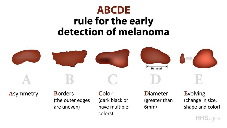
The ABCDE Rule
A classic method in dermatology, the ABCDE rule is a quick guide for assessing moles and lesions. Each letter corresponds to a distinctive characteristic:

- Asymmetry: Non-cancerous moles are typically symmetrical; asymmetry raises suspicion.
- Border: Well-defined borders are characteristic of benign moles. Irregular or undefined borders are cause for concern.
- Color: Benign moles usually have a consistent color. More than one color or shade can indicate potential issues.
- Diameter: Moles larger than 6mm in diameter, approximately the size of a standard pencil eraser, may require further evaluation.
- Evolution: Any changes in size, shape, or color should be monitored closely.
Referral is needed if any of the features appears.
As an example, we notice 4 out of the 5 features on this mole, which is actually melanoma. It is not symmetrical and has an atypical border (not well defined and really irregular). It has several shades of brown and red and is larger than 6 mm. All these signs should alert and need referral.
Both methodologies emphasize the importance of timely referrals if any concerning features are identified. Moreover the major signs of 7PCL are similar to those of ABCDE. Early detection and intervention play pivotal roles in managing potential skin issues!
References
Walter FM, Prevost AT, Vasconcelos J, Hall PN, Burrows NP, Morris HC, Kinmonth AL, Emery JD. Using the 7-point checklist as a diagnostic aid for pigmented skin lesions in general practice: a diagnostic validation study. Br J Gen Pract. 2013 May;63(610):e345-53. doi: 10.3399/bjgp13X667213. PMID: 23643233; PMCID: PMC3635581.